Cell Cycle

Actively dividing cells undergo a series of stages known as the cell cycle.

  • Gap 0 (G0; quiescent phase)
    • Most non-proliferative cells in the body are in this phase and are not actively replicating.
  • Interphase
    • Gap 1 (G1)
      • Metabolic changes prepare the cell for division.  At a certain point (restriction point or G1 checkpoint), the cell is committed to division and moves onto S-phase
    • Synthesis phase (S phase)
      • DNA synthesis replicates the genetic material such that each chromosome is duplicated into two chromatids
    • Gap 2 (G2)
      • Metabolic changes assemble the cytoplasmic materials required for mitosis and cytokinesis
  • Mitotic phase (M phase)
    • Prophase
      • Nuclear membrane breaks down into a number of small vesicles
      • Centrosome duplicates and each one migrates to opposite poles of the cells where they organise production of microtubules that form spindle fibres (mitotic spindle)
      • Chromosomes condense into compact structures.  Sister chromatids are held together via the centromere structure
    • Prometaphase
      • Chromosomes migrate to the middle of the cell (the metaphase plate).
      • Spindle fibres attach to centromeres of the chromosomes.
    • Metaphase
      • Chromosomes align along the metaphase plate
    • Anaphase
      • Centromeres divide and daughter chromatids are pulled apart by spindle fibres
    • Telophase
      • Nuclear membrane reforms around the new chromosomes at either pole of the cell.
      • Chromosomes uncoil and become diffuse once more and spindle fibres are broken down
    • Cytokinesis
      • Constriction of the cytoplasm to divide into two new cells.

Preoperative management of PMHx/Drugs

See also Diabetes and Surgery

Cardiac disease

  • Drugs:
    • Statins can be continued as normal
    • Beta-blockers can be continued (but should not be started if patient was not previously taking them)
    • Antiplatelets should be withheld 7-14 days prior
    • ACE inhibitors and ARBs should be withheld the day of surgery (they can cause marked hypotension with GA)
    • Diuretics should also be withheld on the day of surgery
    • Warfarin should be withheld 3-5 days before surgery (see below)
    • Calcium channel blockers can be continued
  • Pre-operative risk and management
    • Get a cardiology review if there is any concern over the patient’s fitness for surgery
    • For patients undergoing non-cardiac surgery, the ACC/AHA have produced the following guide flow-chart

jjj

    • **Risk**
      • Using the Revised Lee Cardiac Index (RLCI)
        • Any 2 or more of the following would be high risk (>1% risk of major cardiac event)
          • PMHx of MI, (positive ETT, Angina, use of GTN, ECG with pathological Q waves or signs of ischaemia)
          • PMHx of CCF/HF; (pulmonary oedema, PND, bilateral rales or S3 gallop, CXR showing pulmonary vascular redistribution)
          • PMHx of stroke/TIA
          • Preoperative treatment with insulin
          • Preoperative eGFR <30ml/min
    • **METs** (Metabolic equivalent- 1= 3.5ml O2 uptake/kg/min (resting O2 intake))
      • These are similar to assessing someone’s exercise tolerance
        • Self care, eat, dress, toilet etc – 1 MET
        • Walk up a flight of stairs/hill or walk briskly for prolonged time (~4 METs)
        • Can do heavy work, or climb 2 flights of stairs (6-10 METs)
        • Can do strenuous exercise (10+ METs)
  • In patients with unstable Coronary artery disease, it may be appropriate to perform revascularisation (PCI) prior to surgery.  However, this would only represent a minority of patients.
  • Patients with Valvular disease (in particular stenoses) should be considered for peri-operative antibiotic therapy to reduce the risk of endocarditis
  • Post-operatively
    • Make sure to monitor any signs of silent ischaemia (cardiac monitoring) and heart failure

Respiratory Disease

  • The main issue with surgery in patients with respiratory disease is due to anaesthesia
    • Sedation can cause hypoventilation and atelectasis, worsening hypoxaemia and hypercapnia, increased V/Q mismatch
    • Airway manipulation can cause a reactive bronchospasm which can be severe in patients with airways disease
    • Controlled ventilation may cause impaired airflow and increased hyperinflation of the lungs in patients with COPD (and even ‘dynamic hyperinflation’ i.e. continuous inflation of the lungs
    • As such, if possible, avoid general anaesthesia (i.e. use regional anaesthesia)
  • Assessing/managing risk
    • Pulmonary function tests are crucial.  Note that most operations will result in a reduction in pulmonary function peri- and postoperatively, and this should be taken into account when deciding if surgery is appropriate
      • Deep breathing exercises +/- chest physiotherapy/rehabilitation is often useful in patients with COPD to improve function prior to surgery
      • If FEV1/FVC ratio <50%- risk of respiratory failure following surgery is increased dramatically
    • Smoking cessation- this will reduce the risk of post-operative complications including wound healing and pulmonary complications
    • Intra-operative PEEP (positive end expiratory pressure) and post-operative non-invasive ventilation (CPAP or BIPAP) may prevent respiratory failure
    • Make sure to correct any exacerbations prior to surgery
  • Drugs
    • Inhalers/nebulisers should be taken pre-operatively (ideally close to induction)
    • For steroid use, see below
    • Note that anaesthetic drug choice may be important
      • Nitrous oxide may rupture bullae in COPD and cause pneumothorax
      • Opiates usually cause respiratory depression
      • Post operative pain may result in respiratory depression
      • General anaesthesia
        • Reduces muscle tone and thus residual capacity
        • Increases airway resistance and reduces lung compliance
        • Causes atelectasis in dependent zones (causing increased V/Q shunting)
        • Increases ventilatory dead space

Liver Disease

  • Assessment
    • Contraindications to surgery include Acute or fulminant hepatitis, alcoholic hepatitis and severe chronic hepatitis
    • For other patients with liver disease, there are several scoring systems used to categorise risk (Child-Pugh and MELD scores)
      • In general, CP class A/MELD score <10 can undergo elective surgery; CP class B/MELD score 10-15 can undergo elective surgery with caution (see below) and CP class C/MELD score >15 should not undergo elective surgery
      • cp
  • Optimisation
    • In patients with prolong PT- vit K can be given pre-operatively to correct this
    • In patients with ascites and oedema, diuretics may be used to reduce this (alternatively ascites may be drained intraoperatively)
    • Electrolyte abnormalities should be corrected and renal function evaluated/optimised.
    • Patients with gastroesophageal varices should be treated optimally (whether with betablockers/nitrates or with banding/ligation) prior to surgery
    • Where possible, correct any jaundice prior to surgery

Diabetes (see diabetes and surgery)

Thyroid disease

Hypothyroid

  • Potential adverse outcomes
    • Low cardiac output and increased risk of CVD (increased risk of MI; hypotension)
    • Blood loss poorly tolerated
    • Respiratory centre less responsive to O2 and CO2 pressures (hypoventilation; acidosis)
    • More sensitive to opiates
    • Hypothermia
    • Hypoglycaemia
    • Hyponatraemia
  • Management
    • In overt hypothyroidism- correction (levothyroxine) should ideally be given prior to surgery where possible
      • In severe cases (myxoedema coma)- T3 and T4 may be given prior to surgery

Hyperthyroid

  • Increased risk of
    • tachycardia; labile BP and arrhythmias (increased output and contractility due to increase in O2 demand)
    • dyspnoea (similar reason)
    • Thyroid storm- an uncontrolled release of thyroid hormone.  Causes hyperthermia and metabolic acidosis (high mortality)
      • Note that treatment is the same as for hyperthyroidism but increased dose/frequency and adequate ITU support. 
  • Management
    • Ideally controlled with carbimazole or propylthiouracil prior to surgery
      • If surgery is urgent and hyperthyroidism not controlled- potassium iodide drops may temporarily halt to the release of hormones (not temporarily)
    • Propanolol can be used for symptomatic relief

A note about some drugs

  • Steroids
    • Ideally, patients should not be on steroids, as they can lead to
      • Poor wound healing
      • Infection
      • Impaired glucose tolerance
      • Muscle wasting
      • Electrolyte disturbances
      • Masking of sepsis
    • However, patients that are taking or have recently (< 3 months) taken steroids at a dose of >10mg/day are at risk of adrenocorticoid insufficiency should they be stopped.
      • Peri-operatively, this could potentially cause cardiac failure or an Addisonian crisis
      • As such, steroids should be given to cover for this in these patients
      • jjj
        • Dosing equivalents: Prednisolone 10 mg is equivalent to Betamethasone 1,5 mg or Cortisone acetate 50 mg or Dexamethasone 1.5 mg or Hydrocortisone 40 mg or Deflazacort 12 mg or Methylprednisolone 8 mg
  • Warfarin
    • Due to the risk of bleeding, warfarin should ideally be stopped 3-5 days prior to surgery (INR <1.5)
    • If the risk of thrombosis is high (e.g. metallic heart valve); then warfarin should be replaced with heparin.  If the risk is relatively low e.g. AF (without previous CVA), then it may be possible to stop without any heparin substitute.
  • Antiplatelet agents (aspirin, clopidogrel etc)
    • Should be stopped 7-14 days prior to surgery due to risk of bleeding.
  • Anti-epileptics
    • Should be continued where possible

Olfactory Nerve (CN I) and Smell

Background

  • CN I/Olfactory nerve is the shortest cranial nerve and consists of special unmyelinated sensory nerves responsible for sense of smell
    • They are, however, covered in Schwann cells

The olfactory pathway

  • Odourant molecules can enter your system via your nose or mouth and nasopharynx.
  • They reach an area called the olfactory mucosa in the nasal cavity
    • Here, they come into contact with the olfactory receptor neurons and activate receptors to initiate an action potential
      • The axons of these cells traverse the cribiform plate of the ethmoid bone at the roof of the nasal cavity and become the olfactory bulb.
    • In the bulb, these neurons communicate with specialised mitral cells at the synaptic glomeruli.  These then pass posteriorly into the olfactory tract
      • This runs along the inferior aspect of the frontal lobe.  At the anterior perforated substance, the tract divides into medial and lateral stria.
        • Medial stria connects to the limbic system and communicates with the contralateral olfactory medial stria
        • Lateral stria continues to the primary olfactory cortex in the temporal lobe, which goes on to communicate further with the limbic system (amygdala, piriform cortex and olfactory tubercle) and orbitofrontal cortex.
  • Note
    • CN I is covered by pia and arachnoid layers (i.e. continuation of the brain).  It does also not join with the brainstem.
    • The olfactory nerve is capable of regeneration.

Olfactory Dysfunction (Anosmia)

  • Temporary anosmia is not uncommon in local conditions of the nose e.g. infection.
  • Other causes of anosmia/abnormal sensation of smell include
    • Tumours in the olfactory groove (meningioma)
    • Head injury- damage to the cribiform plate may cause damage to the olfactory receptors
    • Neurodegenerative disease e.g. Parkinson’s Disease, Huntington’s and Alzheimer’s disease
    • Genetic conditions e.g. Kallman Syndrome, Primary ciliary dyskinesia, Foster Kennedy Syndrome
    • Partial epilepsy (pre- and post-ictal)

Testing CN I

  • Ask the patient if they have noticed a change in their sense of smell.
  • A more formal assessment of smell can involve using common smells

Slipped Upper Femoral Epiphysis

Background

  • One of the most common adolescent hip problems (around 10/100,000 children per year)
    • The epiphysis usually slips posteriorly relative to the diaphysis of the femur
  • Most common in boys and occurs usually around the growth spurt in adolescence (mean age 13)
    • More common in overweight children; left hip slightly more prevalent
    • Whilst weight and mechanical factors (as well as others e.g. hypothyroidism; hypopituitarism; radiation treatment) may play a role, SUFE represents an underlying instability of the proximal growth plate- the exact cause of which is unknown

Classification

  • The most important classification is whether the joint is
    • Stable (90%) i.e. the patient is still able to weight bear (function relatively unimpaired)
    • Unstable (10%) the patient is unable to weight bear- requires urgent management
  • Other classifications include
    • time-based i.e. acute (symptoms for < 3 weeks); chronic and acute on chronic
    • Southwick angle classification (measurement of the difference between both hips in the femoral head-shaft angle on the frog radiograph)
      • Mild <30°; Moderate 30-50° and severe >50°
    • Grading by degree of slippage
      • I (Up to a third); II (up to a half); III (more than half)

Presentation

  • Most commonly presents with hip and/or groin pain
    • Often an acute event but may present but can have had mild symptoms preceding this
    • Can present as radiated knee pain
    • Worse on movement/weight bearing
    • May cause antalgic gait
    • May limit hip movement- particularly internal rotation and abduction (indeed the leg may rest in external rotation/adduction)
  • Differential
    • Perthes disease
    • Septic arthritis
    • Developmental dysplasia
    • Synovitis

Investigation

  • X-rays
    • An x-ray of the pelvis will usually detect a SUFE
      • Trethowan’s sign
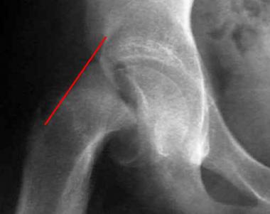
        • Klein’s line (the line drawn up the lateral edge of the neck of the femur) should intersect the femoral head. It fails to do so in SUFE due to slip.
      • You may also see widening of the growth plate (epiphysiolysis) and blurring of the proximal femoral metaphysis (overlapping of the metaphysis and displaced epiphysis)
  • Occasionally, where diagnosis is in doubt, a CT or MRI may help confirm the diagnosis

43288tn

Management

  • Surgical management of the affected side
    • Percutaneous fixation with cannulated screw(s)
  • There is some controversy as to whether to fix the other side also (bilateral in up to 20% of cases)- currently not recommended
  • Without fixation, particularly in unstable cases, there is a risk of osteoarthritis; chondrolysis (breakdown of cartilage and subsequent bony degeration/damage) and avascular necrosis of the femoral head

Scoliosis

Abnormal curvature of the spine in the coronal plane (>10°).

Background/epidemiology

  • Scoliosis <10° is not abnormal (considered a normal variation)
    • Around half of patients with AIS (adolescent idiopathic scoliosis- most common form) develop a curvature >70°
  • It is estimated to affect 2-3% of the population; 80% is idiopathic
  • The most common type is adolescent idiopathic scoliosis (affects between 0.5 and 3% of people, most commonly between the ages 12-14, and, in IAS, is much more common in females (90%)
    • Other common types include
      • Juvenile idiopathic scoliosis (aged between 3 and 10; more common in females; likely to progress/require surgery due to curvature presenting prior to growth spurt at puberty)
      • Infantile idiopathic scoliosis (< age 3; more common in males)
      • Congenital idiopathic scoliosis
      • Neuromuscular and Pathologic Scoliosis (Secondary conditions)
  • In older children, curvature is usually to the right.  In infants, left sided curvature is more common.
  • As curvature progresses, vertebral bodies rotate towards convexity and spinous processes away from convexity.  In severe cases, this can impair cardiorespiratory function.

Presentation

  • May be asymptomatic and detected by chance- note that screening is not routinely offered in the UK (some patients also have a family history)
  • Usually present with back (thoracic) pain
  • On examination (see back examination)
    • Shoulders/waistline may not be level and/or ribs/scapulae may be more prominent in certain areas
    • Note that the hip usually protrudes on the concave side
    • Adam’s Test
      • Ask the patient to bend forward- a fixed scoliosis becomes more prominent
    • It is important also to look for leg length inequality; any focal neurology (change in reflexes) or any signs of congenital/hereditary conditions e.g. midline skin defects, cafe au lait spots.

Investigations

  • XR spine
    • Calculating the Cobb angle (between the uppermost and lowermost vertebra of the primary curvature seen on erect AP XR) is important in deciding management/prognosis

Management

  • Exercises
    • Back exercises have very little effect on curvature but can maintain mobility/range of movement and may improve pain
  • Bracing
    • Used mainly for curvatures between 20° and 40°, which are well balanced (i.e. have a compensatory secondary curve), and in patients who are growing (in puberty) in which a brace may halt the progression and occasionally improve the deformity
    • Usually not definitive- used mainly to maintain curvature stability in younger patients until adolescence when operative management may be more suitable
  • Surgery
    • Spinal fixation (posterior spinal fusion most common) can be used in patients with a curvature of >40°
      • rare but carries risk of neurological complications

Kyphosis and Scheuermann’s disease

Kyphosis is the apical-dorsal curvature of the spine in the sagittal plane (i.e. curves away from the body).  There is normal thoracic kyphosis of around 20-40°.  Abnormal kyphosis is a curvature (measured as Cobb’s angle (between T2-T12)) of >45°.

Background/Aetiology

  • Pathological kyphosis most commonly occurs in the thoracic spine although rarely can occur elsewhere in the spine.
  • It is not uncommon amongst the elderly population (occurs in around 20-30% of patients >65) and is more common in women
  • Risk factors include
    • Osteoporosis; vertebral (wedge) fractures and degenerative disc disease
    • Problems with proprioception; poor posture; spinal muscle weakness/attenuation
    • Other causes include trauma; ankylosing spondylysis and other arthritides; and rarely neoplasms and infections
  • Kyphosis can impair physical functioning; quality of life and mortality directly and indirectly
    • Increased risk of vertebral fracture
    • May affect balance and likelihood of falls
    • Decline in gait speed/mobility
    • In some cases, can impair pulmonary, GI and gynaecological function

Presentation

  • Most patients are asymptomatic and kyphosis is purely aesthetic
  • Some may present with back pain
  • On examination, there is usually a thoracic kyphosis and there may also be compensatory lumber hyperlordosis
    • Tight hamstrings/difficulty with straight leg raise may also be present
  • Rare but important features include those of myelopathy i.e. neurological problems (including pain/altered sensation, weakness, altered tone/reflexes, problems with gait etc)
  • Indications for surgical management (note not absolute)
    • Neurological deficits
    • Kyphosis >70° (no pain) or >65° with pain
    • Loss of anterior vertebral height >50%

Management

  • For purely postural related kyphosis, exercises and education about posture may benefit patient symptoms (may not entirely relieve kyphosis)
  • Surgical management is reserved only for severe cases
    • Smith-Peterson osteotomy, pedicle subtraction and vertebral column resection can all be options for corrections
    • Anterior release spinal fusion may also be used in Scheuermann’s disease/severe cases

Scheuermann’s disease

  • In children and adolescents, abnormal kyphosis can occur as what is thought to be the result of a genetic defect causing collapse of the vertebrae
    • A family history is common
    • Patients
  • Classically defined as anterior wedging of >5° across three consecutive vertebrae, and is different from postural kyphosis by the rigidity of the kyphosis (not corrected by hyperextension)

IgA Nephropathy

Background

  • Also known as Berger’s disease
  • Most common form of idiopathic glomerulonephritis resulting in CKD
    • Around 30-40% of patients go on to develop end-stage CKD within 20 years.
    • Usually presents in young adults and there is a slight male predominance (males tend to have a poorer prognosis)
    • Can be associated with a number of other conditions
      • e.g. Henoch Schonlein purpura; SLE; autoimmune hepatitis; ankylosing spondylitis

Pathophysiology

  • Characterised by IgA- and C3- complex deposition in the glomerular mesangium
  • It is uncertain as to the exact mechanism by which this occurs but patients with IgAN seem to have an raised level of circulating IgA
    • It is thought that a specific type of IgA (galactose-deficient IgA) is responsible
  • As the disease progresses, several features may be seen (all contribute towards a poorer prognosis- collectively known as Oxford classification)
    • Increased mesangial cellularity
    • Segmental glomerulosclerosis
    • Endocapillary hypercellularity
    • Tubular atrophy/interstitial fibrosis

Presentation

  • Usually presents with frank haematuria following an upper respiratory tract infection
    • May also present with microscopic haematuria (more common in older adults) or an AKI
  • Usually this resolves in several days
    • If haematuria persists, there is usually progression to renal failure

Investigation

  • Urinalysis (dipstick)
    • Usually shows protein and blood
  • Urine microscopy (red cells, leukocytes and casts)
  • Urinary protein and creatinine (24 hour-collection)
  • Plasma IgA may be raised in around 50% of cases
  • Renal biopsy will give a definitive diagnosis

Management

  • Manage any hypertension – ACE inhibitors have shown to be beneficial
    • ACEIs plus Angiotensin receptor blockers may have additional benefit
  • Steroids (a 6 months course of prednisolone) is thought to be protective against progression to ESKD
  • Regular monitoring of renal function is important

Polyuria

Urine output of >3 litres per day.  Note that it is usually accompanied by urinary frequency, but that frequency itself may not indicate polyuria (i.e. frequently passing small vs large volumes).

Pathophysiology

  • In general, polyuria can be a result of anything that causes
    • Increased water intake (polydipsia)
    • Decreased ADH (antidiuretic hormone) secretion (central diabetes insipidus)
    • Decreased peripheral ADH sensitivity (nephrogenic diabetes insipidus)
      • ADH promotes water reabsorption in the renal collecting ducts
    • Solute diuresis (most commonly seen in uncontrolled diabetes mellitus- where high glucose concentrations cause a passive diuresis)

Assessment

  • History
    • Define the extent of polyuria and distinguish between urinary frequency i.e. how much urine
    • If polyuria is present, explore this
      • Onset, Duration, Progression, Triggers, Exacerbating/Alleviating factors
      • Associated symptoms- specifically thirst/drinking (polydipsia); weight changes, (also night sweats)
    • Specific things to ask about include
      • Any recent IV fluids/tube feeds; recent catheterisation/urinary obstruction; recent head trauma/surgery or stroke
        • Note patients can be polyuric following urinary obstruction
    • PMHx
      • Diabetes mellitus
      • Psychiatric disorders – on lithium treatment
      • Sickle cell disease
      • Sarcoid/amyloidosis
      • Hyperparathyroidism
      • Hypertension – on diuretics
      • Alcohol and caffeine intake
      • Smoking history
  • Examination
    • General examination of
      • Blood pressure/pulse
      • Weight
      • Mucous membranes (dry?)
      • Skin (dry, pigmented lesions, ulcers/nodules)
    • Neurological/psychiatric exam (doesn’t have to be extensive but may be further explored if there is any suspicion or positive findings
      • e.g. papilloedema; visual fields etc

Red Flags

  • Abrupt onset or onset in children
  • Night sweats, weight loss (particularly where there is a smoking history too)
  • Psychiatric disorder

Investigations

  • Serum or fingerprick (BM) glucose measurement and urinalysis to rule out diabetes mellitus
  • Where hyperglycaemia is absent
    • U&Es including serum Calcium
      • Hypernatraemia suggests excess water loss due to diabetes insipidus
      • Hyponatraemia suggests excess free water intake (polydipsia)
    • Urine osmolarity
      • usually low with water diuresis and high with solute diuresis
  • If a diagnosis is not yet clear, a water deprivation test can be done
    • (NB only to be done as an inpatient under supervision)
      • Typically a baseline set of weight, bloods and observations are performed in the morning.  The patient is then deprived of water.  Where possible, hourly samples of urine are tested for osmolarity and sodium concentrations.  Once the patient shows signs of deprivation (i.e. orthostatic hypotension; postural tachycardia; >=5% weight loss OR if the urinary concentration does not increase by >30mOsm/kg), baseline measurements are repeated and a bolus of exogenous ADH administered.  One hour later, measurements are repeated again.
    • Interpretation
      • Normal
        • Maximal urine osmolarity after dehydration (>700mOsm/kg), and osmolarity does NOT increase >5% following ADH injection
      • Central diabetes insipidus
        • Urine osmolarity unchanged during water deprivation but concentrates following ADH administration
      • Nephrogenic/peripheral diabetes insipidus
        • Urine osmolarity shows no significant change after either water deprivation or ADH administration
      • Psychogenic polydipsia
        • Initial urine osmolarity is low (<100mOsm/kg), but urine concentration will normalise with water deprivation (essentially normal response)
    • Other tests which may be appropriate include
      • Pituitary function tests
      • Serum lithium concentration
      • Autoantibody screen

Causes

polyuria

  • Other causes include
    • Cushing’s syndrome/disease
    • CKD
    • Hypercalcaemia
    • Fanconi’s syndrome

Cardiac Tumours

Epidemiology

  • Primary cardiac tumours are extremely rare
    • Metastatic tumours are 30-40 times more prevalent
    • Of all the primary tumours, myxoma is the most common, accounting for up to half of all primary benign tumours in adults
      • In children, rhabdomyomas are more common

Types

  • Benign Primary tumours
    • Myxoma
      • The majority seem to be located in the left atrium and are pedunculated.  They may prolapse through the mitral valve during diastole and prevent ventricular filling.
      • They can be smooth/firm or friable/irregular.  The latter can often present with systemic embolism.
    • Papillary fibroelastomas
      • Avascular papillomas usually occurring on the mitral or aortic valves.  They don’t often cause any valvular disruption but present instead with embolic disease.  Usually found in older people.
    • Rhabdomyomas
      • Most common heart tumour in children, and commonly associated with Tuberous Sclerosis.  Most are located intramurally or free wall of the left ventricle, and can affect the conduction system.  They can regress with age but a minority can develop tachyarrhythmias/heart failure due to outflow obstruction.
    • Other types of benign tumours include fibromas (often found in childhood and associated with renal tumours or basal cell naevus syndrome); Haemangiomas; Teratomas; Lipomas; Paragangliomas (including phaeochromocytomas) and Pericardial cysts)
  • Malignant tumours
    • Sarcoma
      • 2nd most common primary tumour and most common malignant primary- it affects mainly middle-aged adults and originate in the right atrium, involving the pericardium and can cause right ventricular inflow obstruction and pericardial tamponade.  It frequently metastasises to the lung.
    • Other types include lymphoma and pericardial mesothelioma.
  • Metastases
    • Most common although rare in general- usually from the lung, breast or kidney cancers.

Symptoms/Signs

  • Many patients can be asymptomatic (found incidentally on imaging)
  • Symptoms largely depend on the size, location and character of the tumour, but there may be a triad of features
    • Valvular obstruction
      • I.e. left or right sided heart failure
        • Left sided: shortness of breath; orthopnoea; pulmonary oedema
        • Right sided: peripheral oedema; ascites; raised JVP
    • Embolic events
      • Most tend to be left sided and therefore systemic embolic events
        • Stroke or tissue/organ ischaemia
    • Constitutional symptoms
      • Weight loss, fatigue, weakness, fever, arthralgia
      • May resemble infective endocarditis
  • A diastolic murmur may be audible with myxomas, and an audible ‘tumour plop’ may be heard too as the tumour passes through the mitral valve.

Investigation

  • Due to the rarity of these tumours- often symptoms are investigated for other causes and diagnosis may be delayed
  • Echocardiogram may detect tumours
  • Cardiac MRI is used to stage tumours

Management

  • Depends on the tumour type, individual patient and predicted outcome
  • In general
    • Benign primary tumours should be offered excision (note that this is entirely dependent on individual and their functional status
    • Malignant primary tumours are general palliative
    • Metastatic disease should be treated depending on the primary

Brugada Syndrome

An ECG abnormality with a high incidence of sudden death in patients with structurally normal hearts.

Aetiology

  • Most cases due to a mutation in a cardiac sodium channel gene (sodium channelopathy)
    • Most are spontaneous but occasionally there are hereditary cases

Features/Diagnostic criteria

  • NB Can be transient and/or can be unmasked/augmented by a number of factors e.g. fever, ischaemia, drugs (notably sodium channel blockers e.g. flecainide; calcium channel blockers; alpha receptor agonists; beta blockers; nitrates; cocaine; alcohol), hypokalaemia, hypothermia, cardioversion
  • ECG findings
    • Type 1
      • Coved ST segment elevation >2mm in >1 of V1-V3, followed by a negative T wave (Brugada sign)Stbrugada_(ECG).svg
    • Note that this is the only sign that is potentially diagnostic.  It must be associated with one of the following to make the diagnosis:
      • Episode of VF or polymorphic VT
      • Family history of sudden cardiac death <45 years old
      • Coved type ECGs in family members
      • Inducibility of VT with programmed electrical stimulation
      • Syncope
      • Nocturnal agonal respiration
    • Type 2 sign has >2mm of saddleback shaped ST elevation and type 3 can be either morphology but with <2mm elevation

Other investigations

  • Where the patient is asymptomatic, it may be appropriate to carry out electrophysiological testing to try and induce VT in patients with ECG changes.  However, this is often not conclusive and does carry significant risk.
    • Alternatively, attempting to induce VT with a dose of flecainide may also help with diagnosis, but has similarly poor diagnostic capability and can be dangerous.

Management

  • The only proven management for Brugada syndrome is an ICD device.

ej-vol7n25-fig3